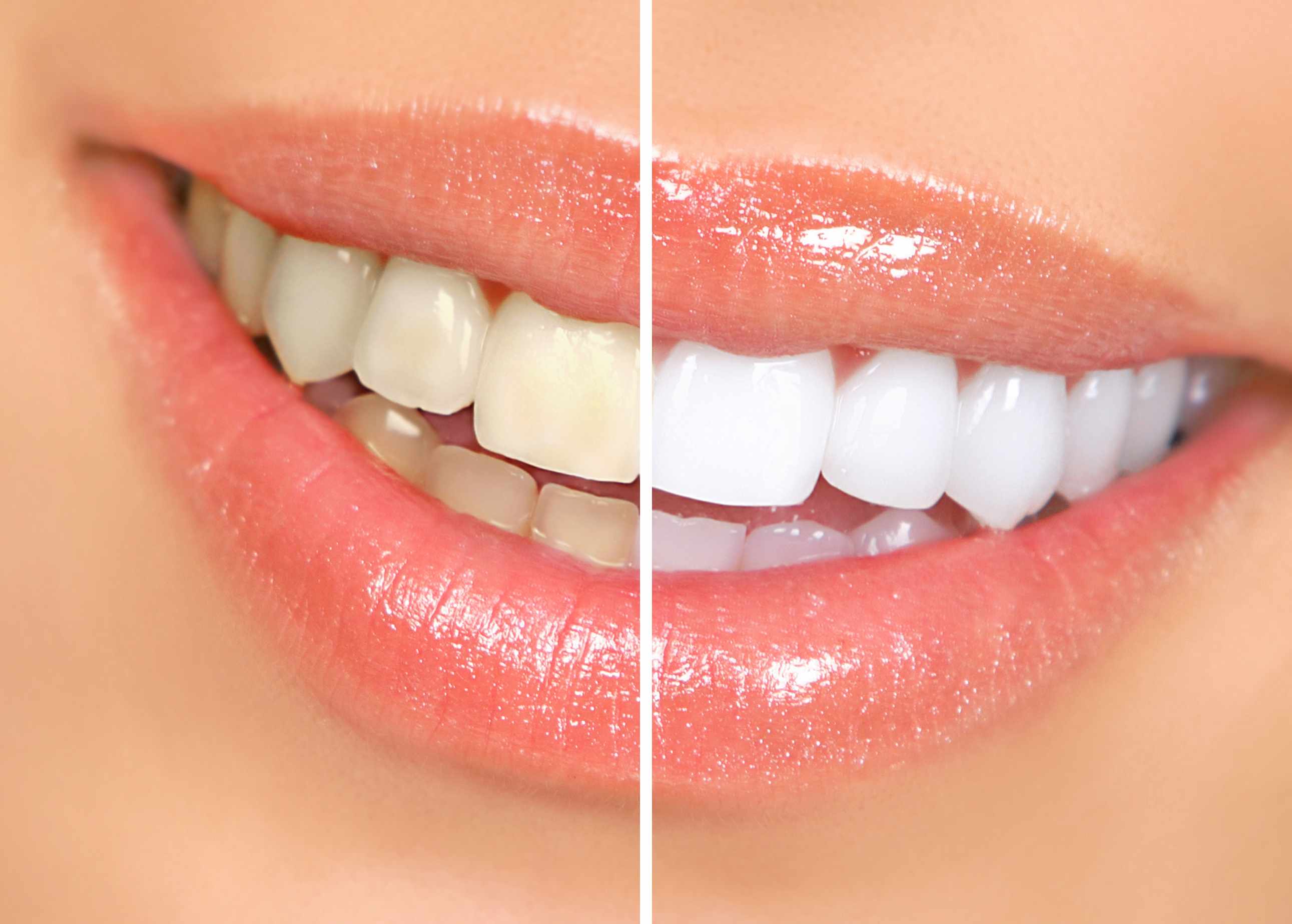
| subject | 치아미백은 바이오필름(치면세균막)에 유리한 환경을 만듭니다! |
|---|---|
| writer | 관리자 |
| date | 19-01-28 11:35 |
| hit | 1,483 |
관련링크본문
치아미백이 치면을 거칠게 해 바이오필름 생성을 쉽게 만든다는 논문 따라서 치아미백 전후 치면을 매끈하게 해주는 트리트먼트가 반드시 필요합니다!
J Dent. 2014 Nov;42(11):1480-6. doi: 10.1016/j.jdent.2014.08.003. Epub 2014 Aug 15.
In-office bleaching gel with 35% hydrogen peroxide enhanced biofilm formation of early colonizing streptococci on human enamel.
Ittatirut S1, Matangkasombut O2, Thanyasrisung P3.
1
Esthetic Restorative and Implant Dentistry Program, Chulalongkorn University, Henri-Dunant Road, Bangkok 10330, Thailand.
2
Department of Microbiology and DRU on Oral Microbiology, Faculty of Dentistry, Chulalongkorn University, Henri-Dunant Road, Bangkok 10330, Thailand. Electronic address: oranart@gmail.com.
3
Department of Microbiology and DRU on Oral Microbiology, Faculty of Dentistry, Chulalongkorn University, Henri-Dunant Road, Bangkok 10330, Thailand. Electronic address: tpanida@gmail.com.
Abstract
OBJECTIVES:
To compare the effects of 25% and 35% hydrogen peroxide in-office bleaching systems on surface roughness and streptococcal biofilm formation on human enamel.
METHODS:
Enamel specimens (3mm×3mm×2mm, n=162) from human permanent teeth were randomly divided into 3 treatment groups (n=54 each): (1) control, (2) bleached with 25% hydrogen peroxide (Zoom2™), and (3) bleached with 35% hydrogen peroxide (Beyond™). The enamel surface roughness was measured by a profilometer before and after treatments. Subsequently, the treated enamel specimens were randomly placed into 3 subgroups (n=18 each) and incubated with: (1) trypticase soy broth control, (2) Streptococcus mutans culture and (3) Streptococcus sanguinis culture for 24h. Biofilm formation was quantified by crystal violet staining. The biofilm structure on three specimens from each group was visualized by scanning electron microscopy. Data were analyzed by Kruskal-Wallis and Mann-Whitney U tests with Bonferroni corrections. Significance level was set at p<0.05.
RESULTS:
Both bleaching systems significantly reduced enamel surface roughness comparing to the control group (p<0.001), but there was no difference between the two treatment groups. Remarkably, S. sanguinis biofilm formation was significantly higher on enamel specimens bleached with 35% hydrogen peroxide than other treatments (p<0.001), but was lower on those bleached with 25% hydrogen peroxide (p<0.001). In contrast, no difference in S. mutans biofilm formation was observed among the three treatment groups.
CONCLUSION:
Both 25% and 35% hydrogen peroxide caused similar degrees of reduction in enamel surface roughness. Nevertheless, bleaching with 35% hydrogen peroxide appeared to markedly promote S. sanguinis biofilm formation.
CLINICAL SIGNIFICANCE:
The increase of early colonizer biofilm raised concerns over adverse effects of in-office bleaching on plaque formation. This should be further investigated in vivo and efficient plaque control should be emphasized after bleaching with high concentrations of hydrogen peroxide.
Copyright © 2014 Elsevier Ltd. All rights reserved.
KEYWORDS:
Biofilm formation; Enamel; Hydrogen peroxide; In-office bleaching; Streptococcus mutans; Streptococcus sanguinis
|
|
댓글목록
등록된 댓글이 없습니다.
